Dermatochalasis — Excess Upper Eyelid Skin
Ageing skin gradually loses collagen and elasticity, and the upper eyelid is one of the areas where this shows earliest and most visibly. Excess skin descends over the lid margin, sometimes far enough to rest on the lashes or genuinely obstruct the upper visual field in more advanced cases. The problem, by definition, is skin quantity — the underlying lid margin and the muscle that lifts it are functioning normally underneath that excess fold. Sun exposure, smoking, significant weight fluctuation and a family history of early skin laxity can all make dermatochalasis appear earlier or more pronounced than typical age-related change alone would explain, though the eventual mechanism — stretched, redundant skin rather than a muscle problem — is the same regardless of cause.
Patients often describe the effect long before they can name it: a heaviness or tiredness around the eyes even after a full night's sleep, difficulty applying eye make-up because there is no longer a visible crease to work with, or a sense that peripheral vision on the outer, upper side has quietly narrowed. Because the change happens gradually over years, many people underestimate how much skin has accumulated until they see an old photograph for comparison, which is one reason a surgeon will typically take standardised photographs at consultation rather than relying on the patient's own sense of "before".
The operation is upper blepharoplasty: the surgeon marks and removes a carefully measured strip of excess skin, sometimes with a small amount of underlying fat if it's contributing to the heaviness, and closes the incision within the natural upper-lid crease so the resulting scar is well hidden once healed. Because the lid margin itself was never the problem, a properly performed blepharoplasty on genuine dermatochalasis typically gives a clean, predictable improvement — the hooding is gone and the eye looks more open, without any change needed to the muscle mechanism underneath. Swelling and bruising in the first one to two weeks are normal and temporary; the crease itself typically settles into its final position over several months as post-surgical swelling fully resolves.
Ptosis — a Drooping Eyelid Margin
Ptosis is a different problem entirely. Here, the lid margin itself — the edge of the eyelid that should sit just below the top of the coloured iris — sits lower than normal, because the levator muscle and its tendon (the mechanism that lifts the lid with every blink and on waking) isn't generating enough lift. This can be present from birth (congenital ptosis), develop gradually with age as the levator tendon stretches or thins (involutional or aponeurotic ptosis, by far the most common adult type), or follow trauma, prior eyelid surgery, contact lens wear, or certain neurological or muscular conditions affecting how the muscle itself contracts.
Clinically, ptosis is usually described by cause as much as by appearance: congenital ptosis reflects a levator muscle that never developed normal strength and is often present from infancy; aponeurotic (involutional) ptosis is the age-related stretching or thinning of the tendon that most adult patients on this page are likely to have; mechanical ptosis results from a lid weighed down by a mass, scarring or swelling rather than a muscle problem itself; and neurogenic or myogenic ptosis reflect an underlying nerve or muscle condition and are comparatively rare but important to rule out, since they can point to a diagnosis well beyond the eyelid. Distinguishing between these matters because the surgical approach, and the realistic outcome, differs between them.
The problem here is mechanical — muscle and tendon, not skin — and no amount of skin removal will correct it. The operation is a ptosis repair, most commonly a levator advancement or a tightening (plication) of the stretched tendon, performed to physically raise the lid margin back toward its normal resting height. This is a fundamentally different operation from blepharoplasty, targeting a different structure, even though both are sometimes loosely described by patients as "fixing a droopy eyelid." Recovery follows a broadly similar timeline to blepharoplasty, but because the surgeon is adjusting muscle tension rather than simply removing tissue, lid height and symmetry are checked particularly closely in the first few post-operative weeks, and a small proportion of patients need a minor adjustment if one side settles slightly higher or lower than the other.
Why Misdiagnosis Leads to Disappointing Results
Consider a patient with both problems together, which is a common combination in older adults. If only the excess skin is removed, the underlying droop of the lid margin is still there once the swelling from surgery settles. The patient looks better in some respects — the heavy "hood" of skin is gone — but the eye itself still sits lower and looks smaller than they had hoped, because the actual cause of that smaller-looking eye, the low lid margin, was never addressed. The right answer for this patient would have been blepharoplasty combined with ptosis repair, planned and consented for together from the outset.
This is, in practice, one of the more common sources of patient dissatisfaction after "standard" upper eyelid surgery — not because the blepharoplasty itself was poorly executed, but because it was the wrong (or incomplete) operation for what was actually present. It underlines why a thorough pre-operative assessment matters more here than almost any other decision in the whole process.
There is also a less obvious failure mode worth knowing about: removing too much skin from an eyelid that has undiagnosed ptosis can occasionally make the lid margin appear to sit even lower once swelling settles, because the skin that was previously providing a small amount of camouflage over the low margin is now gone. This is not a complication of blepharoplasty in the usual sense — the skin removal itself may have been technically fine — but it illustrates why getting the underlying diagnosis right before surgery, rather than correcting it afterwards, gives by far the more predictable and satisfying result for the patient.
How It's Assessed
Standard clinical measurements give an objective picture rather than relying on visual impression alone, and are the basis of the assessment described in general terms by NHS.uk's page on eyelid surgery1:
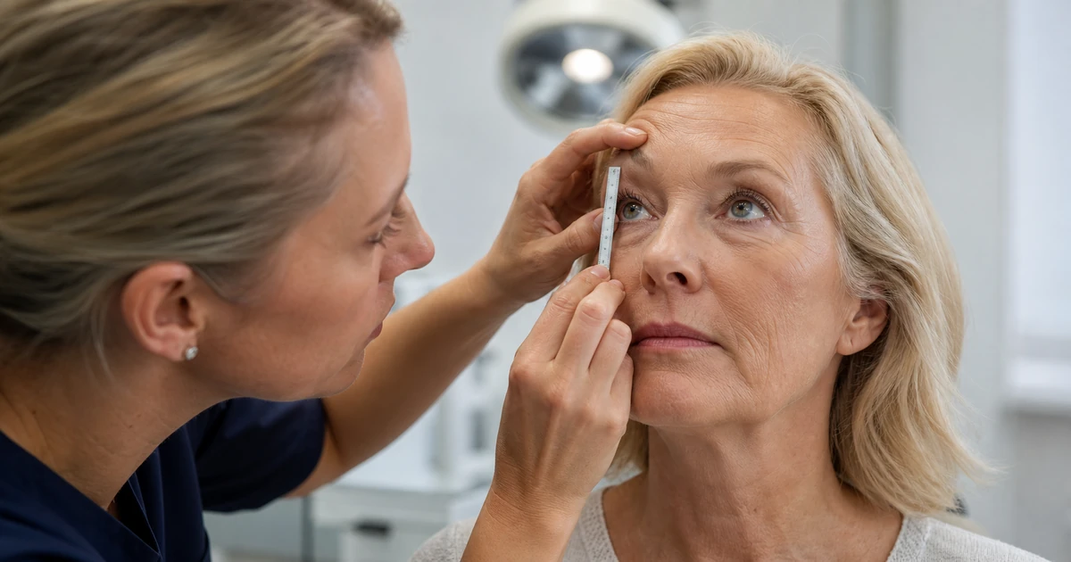
- Margin-reflex distance (MRD1): measured from the corneal light reflex (the small light reflection visible on the front of the eye) to the upper-lid margin. A reduced MRD1 is the key objective sign of ptosis, and the specific number recorded helps grade how significant it is.2
- Levator function: how much the lid moves from full downgaze to full upgaze, measured in millimetres. Good levator function with a low resting lid position points toward a straightforward tightening procedure; poor levator function suggests a more complex underlying muscle problem needing a different surgical approach.
- Skin excess visualisation: a gentle pinch test of the upper-lid skin, sometimes combined with photographs in different gaze positions, to quantify genuinely excess skin separately from the lid-margin position.
- Brow position: assessed at the same time, since a heavy brow can mimic hooding that isn't actually caused by the eyelid at all — see the brow lift guide for how this factor is separately evaluated.
These measurements are quick, non-invasive, and form the basis of a proper surgical plan and consent discussion — a patient should expect to see and understand their own MRD and levator function numbers before agreeing to any specific operation, not simply be told which procedure they need. Standardised photographs are usually taken at the same visit, in primary gaze and with the brow manually held still, since this removes the compensatory brow-raising many patients do unconsciously to keep their eyes open, which can otherwise mask the true severity of either dermatochalasis or ptosis during a casual look in the mirror.
Where both eyes are affected asymmetrically — one lid noticeably lower or heavier than the other — the assessment also needs to check whether the "better" eye is subtly compensating by working harder, since correcting only the more obvious side can sometimes unmask a milder ptosis on the other eye once the stronger side is no longer over-compensating. A surgeon who identifies this possibility before surgery, and discusses it explicitly during consent, is following the more thorough standard of assessment that a two-sided eyelid problem genuinely requires.
Brow Position as a Third Factor
Some "excess upper-lid skin" is actually descended brow position pressing down on the lid from above, rather than true dermatochalasis of the eyelid skin itself. Upper blepharoplasty performed on that patient gives a small improvement — some skin is still removed, after all — but doesn't address the real underlying cause, and the heaviness tends to return, or never fully resolves, because the brow continues to sit low. A thorough assessment should always look at brow, lid skin and lid margin together before recommending blepharoplasty, a brow lift, or both — treating the upper face as one connected system rather than three unrelated areas.
In practice, this means a surgeon assessing "tired-looking eyes" should be manually elevating the brow to its ideal position during examination and observing how much of the hooding disappears purely from that manoeuvre, before concluding how much of the problem is truly eyelid skin versus brow position.
Brow position itself changes with age for reasons distinct from either dermatochalasis or ptosis — the soft-tissue and ligament support that holds the brow up gradually weakens, and the brow descends as a unit, most noticeably at the outer third. A low brow can also make a person look permanently tired, frowning or older than they feel, which is part of why some patients considering upper-eyelid surgery are, on closer assessment, better served by a brow lift instead, by a combined procedure, or occasionally by neither if the brow position turns out to be within a normal range once properly measured. See the brow lift before and after guide for what that separate procedure can and cannot achieve on its own.
Functional Cases, Visual-Field Testing and the NHS
Where skin excess or ptosis genuinely obstructs the field of vision — documented on formal visual-field testing carried out by an ophthalmologist or optometrist, not estimated informally — the NHS may consider funded surgery, because the case is then classed as functional rather than purely cosmetic. NHS.uk on eyelid surgery1 describes the procedure and notes that it is generally private, without setting out the funding pathway in detail; as a general principle across NHS commissioning, specific criteria for functional cases — how much visual-field obstruction qualifies, and what evidence is required — are typically set locally by regional Integrated Care Boards (ICBs) and can vary across England, meaning the same clinical picture may be funded in one area and declined in another.
Patients pursuing an NHS route should expect a formal ophthalmology or oculoplastic referral, visual-field testing as standard evidence, and photographic documentation, usually arranged via a GP referral rather than a direct approach to a cosmetic clinic. Where a case doesn't meet local functional criteria, private treatment remains an option, priced and consented in the usual way.
It is worth being realistic about timescales and outcome on the NHS route: referral, assessment and any subsequent surgery generally take considerably longer than the equivalent private pathway, and NHS funding — where it is granted — covers the functional repair itself rather than any purely cosmetic refinement the patient might also want at the same time. A patient whose case qualifies for functional NHS blepharoplasty should expect the same surgical goal as a private patient (restoring the visual field) but should not expect the operation to be tailored for cosmetic appearance beyond what the functional repair naturally achieves. See Blepharoplasty Cost in the UK for what the private route costs where NHS funding isn't available or isn't wanted.
FAQ
Do I have ptosis or just extra skin?
Look carefully at where the lid margin itself sits relative to your pupil. If the margin is genuinely low, that's ptosis. If the margin sits normally but a fold of skin drapes over it, that's dermatochalasis. Many patients have both together, which only a proper examination can confirm.
Will blepharoplasty fix a droopy eyelid?
Only if the "droop" you're seeing is excess skin (dermatochalasis). A true drooping lid margin is ptosis, caused by the muscle that lifts the eyelid, and needs a separate ptosis repair — blepharoplasty alone will not correct it.
Can the NHS fund it?
Sometimes, for functional cases with documented visual-field loss confirmed on formal testing. Criteria are set locally by each region's Integrated Care Board and vary, so the same case can be funded in one area and declined in another.